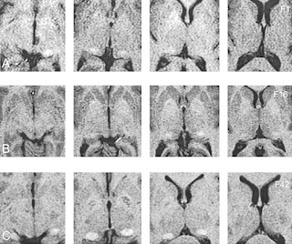
The pulvinus is a part of the thalamus. Isolated pulvinar hyperintensity on T1 weighted MR imaging, thought to be due to calcification, usually bilateral, is thought to be a reliable feature of Fabry disease.
Reference
Moore DF1, Ye F, Schiffmann R, Butman JA. Increased signal intensity in the pulvinar on T1-weighted images: a pathognomonic MR imaging sign of Fabry disease. Am J Neuroradiol. 2003;24:1096-101

Reference
Moore DF1, Ye F, Schiffmann R, Butman JA. Increased signal intensity in the pulvinar on T1-weighted images: a pathognomonic MR imaging sign of Fabry disease. Am J Neuroradiol. 2003;24:1096-101
