Saturday, 18 October 2025

The Man with Septal Perforation

 Cocaine is available in 2 forms- the salt-cocaine hydrochloride, and the "free base", prepared by heating the salt with baking soda. 

The salt can be snorted or injected but cannot be smoked as it disintegrates, while the freebase is smoked. As the freebase is available as white lumps, it is often referred to as "rock". 

When smoked, freebase or rock gives a "high" within 5 minutes. It takes longer for the "high" to appear when snorted (inhaled) through the nose- around 20 minutes.

The man I saw had been snorting cocaine for many years. This is the background against which cocaine induced midline destructive lesion (CIMDL) occurs. Typically, it involves perforation of the septum, but can be more extensive, involve the lateral walls or even the soft or hard palate. 

Such people do not have systemic vasculitis. They have localised destruction of the septum. Systemic symptoms are absent apart from fatigue, which is very common in cocaine users.

These people would typically test positive for pANCA on immunofluorescence, but not for MPO on ELISA. Here, the pANCA is directed not towards MPO, but an antigen called human neutrophil elastase or HNE, considered quite specific for CIMDL.

However, 50% also test positive for c-ANCA and PR3. This causes diagnostic confusion with GPA (Wegener's), which is of course, usually associated with cANCA & PR3 and can cause nasal perforation as well. Unlike CIMDL however, GPA would not show positive pANCA on immunofluorescence. CIMDL patients also tend to have more extensive septal destruction.

A totally different phenotype of cocaine is a genuine vasculitis associated with gangrenous skin lesions and joint pains, alongside systemic symptoms such as fever, night sweats, weight loss, myalgia. A positive urinalysis or proteinuria and lung lesions (the characteristic features of GPA) are only found in a minority of cases. 

The skin lesions are striking- often takes the form of retiform purpura. 

Cocaine abusers with this particular phenotype often have a bizarre double positive on ANCA testing. All of them test positive for MPO ANCA, and around 50% test positive for PR3 ANCA.

It is this latter group, with the genuine vasculitis, that has been associated with levamisole. 

Levamisole is an antihelminth used in veterinary practice, and is used to "cut" (bulk up) cocaine in 70% of such cases. Levamisole has some amphetamine like properties itself and hence ideally suited for abuse.

Cocaine induced vasculitis can be differentiated from GPA through the double positive ANCA, and the bizarre skin lesions. A proportion of such patients are leucopenic, which is rare in GPA. However, just like GPA, cocaine induced vasculitis requires immunosuppression.

Sunday, 1 December 2024

Fasting & Cancer

 Does fasting help cancer? It depends.

Two forms of fasting- one, comprised of eschewing food but taking unrestricted water intake, and the other, called Fasting Mimicking Diet (FMD) have been shown to be useful.

How does it work?

Glucose, amino acids and certain hormones or hormone like substances are thought to encourage cancer growth, including insulin, IGF-1, and leptin. Think of these as anabolic pathways, which provide the nutrients and the drivers for cancer growth and survival. Turn them off, and the cancer cells are disdavantaged.

Note that I haven't mentioned fatty acids, non-intuitive though it may seem. Cancer cells are highly dependent on anaerobic metabolism, epitomised by the Warburg effect, which involves hijacking the glycolytic pathway (normally anaerobic) even in the presence of oxygen, and producing ATP therefrom. This is seen in fully 70-80% of cancer cells.

Conversely fatty acids are metabolised inside mitochondria by beta-oxidation and therefore is a highly aerobic process, not normally utilised by cancer cells.

The corollary is that cancer fighting diets must contain very little carb or protein and any calories, limited albeit in amount, must come from fat. This is exactly what happens with FMD, which consists of 300-1100 calories perday, derived from broths, soups, juices and nutty bars, with some herbal teas thrown in for taste.

It is thought that by abrogating the anabolic hormones mentioned above, you shut down two key canonical intracellular pathways- the PI3K-AKT-MTOR pathhway and the cAMP-Protein Kinase A pathway. Both are proliferatogenic and indispensable for cancer cells.

However, the claim that FMD alone will cure cancer is inaccurate. This diet works well in tandem with chemotherapy and radiotherapy, and must be administered during the peri-chemotherapy period, usually 48 hours before and 24 hours after. It stops the cancer cells from finding "escape" pathways to circumvent chemotherapeutic agents. But the piece-de-resistance of such diets is that it protects normal, healthy cells from being destroyed by chemo, while the cancer cells perish. 

That is to say, the normal cells display Differential Stress Resistance (DSR)  to cancer cells. At the onset of fasting, normal, non-cancerous cells go into a sort of hibernant, low metabolic state, which reduces their vulnerability to chemotherapeutic agents. As they are no longer actively taking up nutients, metabolising or dividing, they become relatively immune to chemo and radiotherapy.

You might well ask- does this work for all cancers? 

Unfortunately not. It's particularly effective for breast cancer, which is ER/PR positive, but not for ER/PR negative,  HER-2 positive cases. Thus, it potentiates the action of both Tamoxifen, without causing endomtrial hypertrophy, and Fulvestrant.

Similar benefits are seen for prostate and colon cancer.

If a relatively fat rich diet works, what about the ketogenic diet (4:1 fat:carb+protein in terms of weight)? After all, the blood ketone levels can rise by >0.5 mmol/L with FMDs.

Neurosurgeons here will know that ketogenic diets have considerable benefits for some intractable childhood epilepsies and has been advocated for certain gliomas/glioblastomas. However, in general, ketogenic diet does not work for cancers, and can worsen prognosis in melanomas. Hence, best avoided.

A few caveats. Most of the data for these findings were from animal, mainly murine studies and from yeasts. Human data is limited.

Secondly, some cancer patients are cachexic at treatment. They would be at risk from FMD like diets.

Thirdly, trying to achieve these metabolic benefits pharmacologically does not seem to work. The MTOR inhibitor Rapamycin (Sirolimus) is not generally useful for cancers.

Fourth, chronic calory restricted diets don't seem to work for cancer. FMD must coincide with the chemotherapy cycles. This is therefore intermittent fasting.


Saturday, 7 September 2024

2024 ACR/ACCP Guidelines on Treatment of SARD-ILDs

The ACR/ACCP committee looked at ILD associated with five common SARD categories:

1. Systemic Sclerosis (SSc)
2. RA
3. IIM (includes anti-synthetase syndromes and Immune mediated necrotising myositis).
4. Sjogren's
5. MCTD

Following is a summary of the recommendations:

For initiation of treatment:

1. MMF as first line choice to commence treatment in all categories. However, this may be tempered by extra-pulonary considerations, for example, rituximab may be preferred in RA with active joint disease.
2. Cyclophosphamide still among first line choices, but Rituximab preferred in all 5 categories due to equivalent results and lower incidence of side effects.
3. Steroids, either short term or long term, to be avoided in SSc.
4. Short term steroids acceptable in other categories, but not long term.
5. Tocilizumab, elevated to one of the first line choices in SSc and MCTD with SSc features.
6. In RA-ILD, Methotrexate, Leflunomide, anti-TNFs and Abatacept should not be used for treatment of ILD. There is discretion to use them for arthritis, but some panelists would withdraw them if incident ILD develops.
7. JAK inhibitors can be used as first line in IIM, and particularly useful in MDA-5 (JAKi include Tofacitinib, Baricitinib and Upadacitinib).
8. Similarly, calcineurin inhibitors, with tacrolimus preferred to ciclosporin, is amongst first line choices for IIM-ILD.
9. Nintedanib is now recommended as a first line choice in  SSc, but not others.
10. Azathioprine remains among first line choices for all categories, but is down in the pecking order for SSc, behind MMF, Rituximab and Tocilizumab.

For progression of ILD despite treatment:

1. If any of the first line Rx above has not been used, it may now be used, with the exception of Azathioprine.
2. Nintedanib can be used for all categories, on the grounds of PPF.
3. Tocilizumab may be used for progressive RA-ILD.
4. IVIG may be used for progressive IIM-ILD.
5. AHST may be considered at this point for SSc-ILD.
6. Pirfenidone may be considered, but only for progressive RA-ILD.
7. Lung transplant can be considered in all categories.

For rapidly progressive ILD (RP-ILD), as seen, for example in MDA-5:

1. IV methylprednisolone, 1g for 3 days, plus at least 2 others from Rituximab, Cyclophosphamide, IVIg, MMF, JAKi and Tacrolimus
2. For non MDA-5 conditions, it is IV methylprednisolone and at least one, or sometimes two of the others.
3. Again, avoid steroids in SSc-ILD.
4. Rituximab and Cyclophosphamide preferred ahead of MMF and calcineurin inhibitors in RP-ILD.
5. Rituximab preferred ahead of Cyclophosphamide in MDA-5 associated RP-ILD.
6. JAKi useful for slowly progressive MDA-5 but not for RP-ILD.
6. Consider early referral for lung transplant in all eligible cases of RP-ILD.

https://acrjournals.onlinelibrary.wiley.com/doi/epdf/10.1002/art.42861?af=R

Thursday, 27 April 2023

The useful Sodium Thiosulfate


Sodium thiosulfate is not the first drug you think of when asked to reel off a series of medications. Yet, it is a versatile, underappreciated agent that can on occasions be lifesaving.

One of its foremost roles is in cyanide poisoning. When you think of cyanide, you invariably conjure up images of the Agatha Christie type pill in the cup of tea type scenario. Yet, that sort of cyanide, usually KCN, is rarely available, and most cases of cyanide poisoning occur to due to smoke inhalation in people caught in fires, from hydrogen cyanide. Here, the three most useful agents are sodium thiosulfate, sodium nitrite and good, old-fashioned vitamin B12- hydroxocobalamin.

You may have heard of cisplatin induced hearing loss? This is particularly problem in paediatric oncology, where IV cisplatin is given to treat a hepatoblastoma, for example. Yet, by limiting cisplatin infusion time to 6 hours, and giving IV sodium thiosulfate within 6 hours of cisplatin, you can reduce the incidence of cisplatin induced hearing loss in these children.

Sodium thiosulfate is particularly useful where vascular calcification leads to troublesome complications such as skin ulcerations in fatty areas in patients with advanced CKD, known as calciphylaxis, or where a rare autosomal recessive disorder called ACDC exists.

ACDC is admittedly rare. It is an inherited, autosomal recessive disorder where relatively young subjects in their 40s and 50s present with severe limb claudication, ischaemic ulcers, critical limb claudication & joint pains in hands and feet. The hallmark is calcification of arteries such as iliac, femoral and tibial arteries, but sparing the aorta, which is very unlike atherosclerosis, which ACDC is often confused with.

ACDC stands for “arterial calcification with deletion of CD73”. You may remember that CD73 converts cyclic AMP to adenosine. Apparently, this leads to higher levels of tissue non-specific alkaline phosphatase downstream, and ectopic calcification. Apart from arterial calcification, you also get periarticular calcification, which can be a clue.

Again, IV sodium thiosulfate in such subjects can be limb sparing.

Saturday, 11 February 2023

Preventing Gentamicin Induced Permanent Deafness in Newborn Children

 You ever heard of a child going deaf after a single dose of Gentamicin? It's more common than you think and it happens regrettably too late to do anything about it, as a single dose of gentamicin can cause permanent deafness in these children.


It happens in 1250 newborns every year in the United Kingdom.


These unfortunate children are 0.2 of the population, who carry a specific mitochondrial DNA mutation.


Remember, mitochondria have a circular DNA comprised of 16,569 base pairs (as opposed to the linear nuclear DNA, which is comprised of 3.3 billion base pairs). The mitochondrial DNA contains 37 genes, which code for 13 proteins, 22 tRNAs, and 2 ribosomal RNAs.


One of those two rRNAs is coded by a mitichondrial gene called MT-RNR1. If a child has a single nucleotide polymorphism, m.1555A>G (which means at the 1555th mitochondrial base pair, adenine has been replaced by guanine), he or she is exquisitely sensitive to the ototoxic effects of Gentamicin, and can develop permanent and catastrophic deafness after a single dose of gentamicin.


This is clearly a problem, as the UK, like many other countries, has guidelines which recommend that the empirical treatment of neonatal sepsis should be with a combination of gentamicin and benzylpenicillin. Moreover, NICE guidance also stipulate that any suspected sepsis must be treated within 1 hour of diagnosis, given its usually severe consequences.


But how do you test for a mitochondrial mutation within 1 hour? Standard genetic tests take hopelessly long.


Well, it seems that the pandemic has tought us a trick or two. At the Manchester Centre for Genomic Medicine, they have developed a point of care test (POCT) using the same technique used for COVID19- PCR amplification- that gives a result within 26 minutes of taking a gum swab from a newborn.


Those testing positive on the POCT are then confirmed with standard (old fashioned) Sanger sequencing and receive Cefotaxime rather than the combination of gentamicin and benzylpenicillin. No deafness!


And how does gentamicin cause deafness in this children? Well, gentamicin kills bacteria by binding to their 16S rRNA and preventing bacterial protein synthesis. In children who have the m1555A>G mutation in their MT-RNR1 mitochondrial gene, the normally eucaryotic 12S rRNA assumes a shape like the bacterial 16S rRNA, and thus binds to gentamicin.


The test has been provisionally approved by NICE, and is currently being piloted prior to a national rollout.

Reference:

AMA Pediatr. 2022;176(5):486-492. doi:10.1001/jamapediatrics.2022.0187

Saturday, 10 September 2022

The Man With Recurrent Leg Swelling

 Saw an interesting case last week.

Thirty-two year old Caucasian man, works loading-unloading shlelves in a supermarket. Over several months, has had recurrent episodes of swelling in his right lower foot, extending from shin, across the ankle into the dorsum of foot. During the episodes, which last several hours, the leg feels very swollen, tight and uncomfortable but not much actual pain.

As usual GP thought of gout, checked urate levels- plumb normal. Does a MRI of the ankle after a normal X-ray. That just shows subcut fluid, joint looks fine. Inflammatory markers normal.

Chap comes to Rheumatology clinic. I get a bit more history. Turns out that the episodes always happen after lunch when he goes back to his rather manual role at the supermarket. He usually has various types of sandwich for lunch.

Has he ever had anything else that seemed unusual? Turns out that in the past, on several occasions, when he had a can of Fosters (a type of lager), his throat felt swollen "inside", so that he would struggle to swallow for hours afterwards, although there was no shortness of breath. He now avoids lager.

The penny drops. The guy has a fascinating condition called "Food Dependent, Exercise Induced Anaphylaxis" (FDEIA). Most published cases in the literature have been in conjunction with wheat consumption, followed shortly afterwards by exercise. Except that now we realise that it's not just exercise, but alcohol and NSAIDs that can trigger this after subjects have fed.

Although in most cases, the putative food contains wheat, other allergens that can trigger it are celery, mustard, fenugreek and coriander. Celery and mustard are of course widely added to spice up pizzas and other takeaways and therefore may be hidden.

You might have guessed that the episodes of "swelling" inside the throat and the leg, both represent angioedema, the first mucosal, the second cutaneous.

This is now called augmentation factor anaphylaxis or cofactor associated food allergy. Avoidance works best, but of course, sometimes the specific food trigger can be hidden, and therefore difficult to identify.

Interestingly celery interacts with birch pollen and a couple of other pollen to give rise to what is described as "Pollen Food Allergy". These people with apparent hay fever, get anaphylaxis type episodes every time they ingest celery.

Such people sgould avoid food that contain a very high concentration of fruit pulp such as smoothies, as the latter is apparently rich in something called Lipid Transfer Protein (LTP), which is very allergenic.

They should also consider carrying an EpiPen, just in case...

Saturday, 5 February 2022

Mycoplasma hominis & Ureaplasma urealyticum Cause Septic Arthritis in Subjects with Hypogammaglobulinemia

 If you have a subject with hypogamma, either due to CVID or other "constitutional" causes, such XLA, or acquired, due to immunosuppression, usually with Rituximab, who present with a picture of inflammatory arthritis, either monoarthritis or oligoarthritis, suspect infection by either Mycoplasma hominis or Ureaplasma urealyticum, particularly if usual cultures are negative and the usual antibiotics do not work.

Both organisms are overrepresented in subjects with septic arthritis developing against a background of hypogamma, particularly on Rituximab. The most commonly affected joints are hip, knee, shoulder, elbow, ankle and PIP. Prosthetic joint infections, particularly affecting the hip have been reported several times.

These fastidious organisms will not grow on routine cultures. M.hominis may show up on anaerobic culture plates after 5 days, but is best cultured on PPLO (pleuropneumonia like organism) broth or looked for by 16S RNA. Similarly, U.urealyticum is best cultured on Ureaplasma differential agar, Ureaplasma broth, or A7B agar. Fortunately, the last 3 will also grow M.hominis. U.urealyticum can also be identified on 16S RNA analysis.

If a person with hypogamma and putative inflammatory arthritis has proven culture negative on synovial fluid and blood culture, do ask for these special media or 16S RNA to isolate these 2 organisms. 

Ureaplasma requires urea in its culture medium for growth. It produces urease with breaks down urea into ammonia, and can thus change the colour of phenol red to pink. M.hominis metabolises arginine, but not glucose. Ureaplasma ferments neither, but produces a "managanese reaction" in culture.

If the usual antibiotics have not worked, there would be a natural tendency to attribute the inflamed joint(s) to the underlying disorder such as RA or Lupus, or make a new diagnosis of one of the above two, and treat with steroids. This may lead to a worsening of such arthritis, so special caution is warranted in subjects with hypogamma.

A course of doxycycline may be indicated if the usual antibiotics which target cell walls (which these organisms lack) have not worked. Resolution of fever/arthritis with doxycycline supports the likelihood of infection by one of the above two.

Tuesday, 21 December 2021

Beware Adult Onset CGD in Subjects with Pulmonary or Extrapulmonary Granulomas but Negative AFB & Tuberculous Culture

 Chronic Granulomatous Disease (CGD) is rare and affects 1 in 200,000 live births. While 75% have the X-linked form (and thus presenting in boys) due to a protein subunit of NADPH oxidase called gp91phox, the rest have an autosomal recessive form due to other protein subunits of the same enzyme, namely p22phox, p47phox, and p67 phox. Of these, p47phox is the second commonest and can present during adulthood occasionally. The involved gene is that of NCF-1 (Neutrophil Cytosolic Factor-1). The average age for presentation of the X-linked form is 3 years, and for the autosomal recessive forms, 8 years.

When CGD presents in adulthood, it is often confused with tuberculosis due to a pulmonary involvement (the most commonly affected organ in CGD), and presence of granulomas on biopsy. These granulomas are however, non-necrotising, and obviously without detectable AFB and yield negative cultures for M.tuberculosis. They are therefore sometimes diagnosed as pulmonary sarcoidosis, given the geographical setting and demographics.

Sputum or bronchoscopic washings growing the following 6 genus in a subject with pulmonary granuloma should lead to a suspicion of CGD- Aspergillus, Candida, Staphylococcus, Serratia, Burkholderia or Nocardia.

These patients often have hyperglobulinemia, with raised Ig levels, but this is not invariable. 

CGD can affect other areas, and thus cause abscesses or cellulitis in the skin, gingivitis (but not periodontitis, unlike leucocyte adhesion defects) Crohn's like granulomas in the intestines, and spinal abscesses. Granulomas can cause obstructive lesions in the urogenital & GI tracts. Delayed wound healing is often a notable feature.

Unlike LAD, which can also rarely present in adulthood, the neutrophil count tends to be normal between infectious episodes. Just for context, there are two forms of LAD, including LAD-1, caused by a defect of CD18, which is a part of the heterodimeric beta-integrins, and LAD-2, caused by a defect in fucosylation, and thus an absence of Sialyl Lewis-X , the latter being necessary for neutrophils to roll on endothelial cells prior to adhesion and diapedesis. Subjects with LAD tend to have severe periodontitis and perpetually high neutrophil counts. LAD-2 is associated with developmental abnormalities such as stunted growth.

The most convenient diagnostic test for CGD is flow cytometry with Dihydrorhodamine 123. Absence of fluorescent staining indicates abrogation of oxidative burst in neutrophils, which is characteristic of CGD. An alternative test is the Nitroblue tetrazolium test (NBT).

The only curative treatment is allogeneic BMT. Symptoms can be improved by treatment with gamma-interferon, and prophylactic administration of co-trimoxazole and itraconazole. Infective episodes should be treated with specific antibiotics.

Sunday, 5 December 2021

Why Extensive Training May Not Help You Run Faster

Practice makes perfect. Right? Not always, it would seem.

I have often wondered why, despite running daily, I have not been able to improve my times. I would be classed as middle to long distance runner, averaging 6 miles daily during weekdays and 9.5 miles on weekends. In the spring of 2020, I thought I had hit a sweet spot, consistently running the longer weekend distance at between 73 and 75 minutes. At 54.5 years of age, that was a reasonable time.

But things went backwards in the autumn of that year, and this year I have struggled to get below 90 minutes for the 9.5 mile run.  In fact most runs have been in the mid to high 90s, with a few taking longer than 100 minutes.

Is this muscle fatigue, perhaps an inevitable consequence of the exercise addict's need to run daily? Or is it due to glycogen depletion?

The answer, it would seem lies elsewhere.

There are two broad categories of muscle fibres, the red, myoglobin rich, largely aerobic, mitochondria laden "slow" Type I fibre, and the larger, predominantly anaerobic, white, glycogen loaded "fast" Type II fibre, that predominantly use glycogen to generate its ATP. 

Type II fibres are further subdivided into Type IIA & Type IIB.

It seems that the determinant of the type of muscle fibre is the myosin heavy chain (MHC, not to be confused with Major Histcompatibility Complex) isotype. Thus Type I fibres are rich in MHC 1, type IIA in MHC 2A, and Type IIB in MHC 2X, also called MHC 2D.

You may well wonder what happened to MHC 2B and 2C? Well, MHC 2B is found in some ultrafast Type II fibres, mainly in cranial muscles, and MHC 2C is found in regenerating muscle fibres.

Biochemically, type I and type II fibres are distinguished by their ATPase staining and the speed of the sarcoplasmic reticulum (SR) in releasing calcium. Thus, Type I fibres stain for ATPase at low pH, while Type II fibres do so at an alkaline pH. Similarly Type II fibres possess SR that release Ca very quickly, while with Type I fibres, the release of Ca from SR is slower.

The distinction between Type IIA and Type IIB fibres is more than that of semantics. Type IIA has characteristics that are intermediate between Type I and Type IIB. Thus, they are versatile in being able to function as slow muscle fibres that serve sustained activity such as gaze, while, when called upon, they can also serve up the explosive speed of contraction, sustainable only for short periods, that epitomises Type IIB fibres. 

Muscle fibres can be "pure" and contain just MHC1 or 2A or 2X, but they can also be hybrid- ie. contain MHC1 & 2A, or 2A&2X, or all three, ie. MHC1, 2A &2X. 

Using immunohistochemical staining, long distance elite runners have a higher proportion of  MHC1 fibres than non-runners, roughly equivalent amounts of MHC2A, and almost no MHC2X fibres. Similarly, non-runners tend to have more hybrid MHC fibres in general than runners, which suggests that hybrid fibres can change their composition depending on the workload imposed on them.

Type I fibres are high maintenance. They tend to be smaller, have larger blood supply and higher oxygen consumption. Type II fibres are larger, but consume low oxygen, and at least the IIB fibres are relatively less used, "standing by" for the relatively less frequent occasions when explosive power is required.

A muscle fibre owes its "type" or allegiance to the alpha motor neuron that innervates it. Thus a motor unit (comprised of all the muscle fibres innervated by a particular motor neurone) will always innervate the same type of muscle fibres. Thus a motor unit can be classified as Fast twitch (F) or Slow twitch (S). The F units are further classified as Fatigue resistant (FR) or Fast fatiguing (FF). When there is denervation and cross re-enervation, the muscle fibres take on the characteristics of the "new" motor neurone. Thus S units can turn into FR or FF on cross-reinnervation and so on.

It follows from the above that with training, entire motor units change their character (as individual muscle fibres are unable to do so, being dictated by the motor unit they are part of). Thus, with prolonged training, FF units can change into FR and FR into S. Change in the other direction happens with deconditioning.

And herein, I believe lies my predicament. I believe that my FF units have slowly transitioned into FR and FR units into S units. Glycogen depletion would not be germane to this, as it would only affect the FF motor units, which are comprised principally of Type IIB muscle fibres. While the S units and FR units are fatigue resistant and ideal for long distance running, the muscle fibres they innervate are smaller, and lack the speed of FF units.

Wednesday, 1 September 2021

Not All Inherited Neurological Disorders are Untreatable

Hereditary Sensory and Autonomic Neuropathy Type 1 responds to large doses of L-serine. It is the only member of the HSAN group that has an effective pharmacologic treatment.

There are a number of inherited and metabolic disorders that respond to large doses of vitamins. These mostly present in adolescents and young adults but can occasionally present later in life.

Thus, a group of 4 disorders respond to large doses of riboflavin. These include 1. Multiple acyl CoA dehydrogenase deficiency, caused by a deficiency of one of 3 enzymes- Electron Transfer Flavoprotein A or B, and ETF dehydrogenase, also called ETF ubiquinone oxidoreductase, responsible for fully 97% of MADD cases. 2. Riboflavin Responsive Exercise Intolerance, caused by a deficiency of mitochondrial FAD transporter, 3. Lipid Storage Myopathy due to deficiency of FAD synthase, and 4. Acyl CoA dehydrogenase 9, or ACAD9 deficiency. All four present with muscle pain, exercise intolerance and a rise in plasma acylcarnitine level of all chain lengths. Episodes of stress associated rhabdomyolysis may occur.

There are two other riboflavin associated disorders: Riboflavin transporter deficiency (RTD) presents as a motor neuronopathy with bulbar involvement, mimicking MND, but can also cause optic and auditory neuropathy. CMTX4 is due to mutations in Apoptosis inducing factor mitochondria associated 1 gene and presents with the classic Charcot Marie Tooth phenotype of hereditary sensorimotor neuropathy. areflexia, pes cavus and additionally auditory neuropathy. Both RTD and CMTX4 show a delayed response to riboflavin supplementation.

Similarly, a number of inherited disorders respond to thiamine. Biotin-thiamine responsive encephalopathy (BTRE)  presents with Leigh syndrome- encephalopathy with T2 changes on MRI in basal ganglia, as does E1 alpha pyruvate dehydrogenase deficiency. In the latter, serum and CSF pyruvate and lactate are elevated in equal proportions.

Biotinidase deficiency presents with a neuromyelitic optica phenotype, comprising bilateral visual loss in combination with longitudinally extensive myelopathy. The disorder responds to oral biotin, which should be started ASAP.

Vitamin B12 deficiency and folate deficiency are probably the most frequently diagnosed vitamin deficiencies in clinical practice. They tend to present with similar phenotypes- gait difficulties, peripheral neuropathy, posterior column involvement and psychosis or dementia, with or without macrocytic anaemia.

It is not surprising therefore that inherited defects of B12 or folate pathways would present with similar manifestations. This can occur at any level from defects of absorption from the gut to deficiency of enzymes that produce the active forms of these vitamins. Thus, in Immerslund-Grasbeck syndrome, mutations in the small bowel mucosal transporter called Cubam, leads to megaloblastic anaemia and neurologic manifestations. Cubam consists of 2 subunits- Cubilin, which recognises the B12-IF complex, and Amnionless, which endocytoses them. Thus, defects in either of the putative genes CUB or AMN can lead to IGS.

B12 serves as a co-factor for 2 important reactions in the cell. Firstly, within the mitochondrion, as S-adenosyl cobalamin, it serves as a cofactor to convert methyl-malonyl Co-A to succinyl Co-A, catalysed by methyl-malonyl Co-A mutase. Succinyl Co-A then enters the TCA cycle. 

In the cytosol, B12, as Methyl cobalamin, is a methyl donor for the conversion of homocysteine to methionine, catalysed by methionine synthase. Methyl cobalamin is regenerated by donation of a methyl group from 5-methyl tetrahydrofolate, which latter itself becomes demethylated to tetrahydrofolate. THF spontaneously converts to 5,10 methylene tetrahydrofolate, which is then reduced by 5,10 methylene THF reductase to the active methyl donor 5-nethyl THF, thus completing the cycle.

Folate is also a cofactor in the latter reaction, but not the former. Thus both B12 and folate deficiency leads to a rise in serum homocysteine, but only B12 deficiency elevates methyl-malonic acid levels.

The B12/folate pathways overlap with the active form- pyridoxal phosphate (PLP)- of another vitamin- Pyridoxine, or Vitamin B6. Thus PLP is a cofactor for the enzyme cystathionine-beta synthase, which converts homocysteine into cystathionine, and eventually into cysteine. Deficiency of this enzyme leads to the hereditary condition homocystinuria, which has some features of Marfan syndrome. In homocystinuria, serum homocysteine levels and methionine levels are both elevated, unlike in deficiencies of the B12-folate pathway, where homocysteine is elevated but methionine levels are low.

Pyridoxal phosphate activity can also be disrupted by inborn defects of two other amino acid pathways- lysine and proline. In the first instance, deficiency of alpha-amino adipic acid delta-semialdehyde dehydrogenase results in conversion of that lysine metabolite to 6-piperideine carboxylate (P6C). In the second case, a condition called Hyperprolinemia Type II results from the deficiency of 5-Pyrroline carboxylate (P5C) dehydrogenase, leading to elevated levels of  P5C. P6C and P5C are both inhibitors of pyridoxal phosphate and thus can again lead to hyper-homocysteinemia.

Disorders of Vitamin E metabolism can lead to two ataxic disorders- Abetalipoproteinemia and Ataxia due to Vitamin E deficiency. Both conditions present with sensory neuronopathy. The former also features retinopathy while the latter features a tremor of the head. The former is due to deficiency of Microsomal triglyceride transfer protein (MTTP) while the latter is due to the deficiency of alpha-tocopherol transfer protein (TTPA) caused by involvement of the putative genes. These two disorders should be considered for all cases of chronic cerebellar ataxia or sensory neuronopathy, which latter presents as non-length dependent sensory neuropathy affecting thighs, arms, etc.


Monday, 15 March 2021

Usefulness of IgG Avidity to Diagnose Recent CMV Infection

 Primary CMV infection acquired during pregnancy is associated with a risk of foetal anomalies such as visual loss, sensorineural deafness, and cognitive impairment. Primary CMV infection is often asymptomatic, hence many women may not know they have been infected. Such infections are particularly common in women working with children such as primary school teachers and day care nurseries.

The prevalence of CMV infections rises with age. Subjects can be re-infected with a different strain or have re-activation of the original CMV strain- CMV is after all a herpesvirus and therefore establishes latency, just like EBV or HZV. It is important to clarify here that the risk of foetal anomalies only arises for primary CMV infections acquired during pregnancy rather than re-infections or re-activation of the virus. CMV infection picked up even shortly before pregnancy do not affect the foetus.

Why should this be so? It is thought that existing antibodies to CMV following a previous infection offer protection against the virus being passed on to the foetus. Thus, longstanding maternal antibodies against CMV bind the viral antigens with a high avidity and protect the foetus.

However, this does not apply for primary infections acquired during pregnancy. Antibodies formed following a recent infection initially bind the viral antigens with low avidity for the first 3-4 months. After this period, due to a process of B-cells with higher antigen specificity being positively selected, a process called affinity maturation, the avidity of CMV antibodies for the virus increases.

This differential avidity is used as an index (Avidity Index or AI) for the recency of CMV infection. Thus low avidity of IgG antibodies for a CMV viral lysate indicates a primary CMV infection within the last 3-4 months, while high avidity indicates one of 3 possibilities- either that the primary infection occurred more than 6 months ago, or that this was a re-activation of a previous primary infection, or that this is a new infection with a different strain of CMV.

The reason this metric is used to gauge the recency of CMV infection is because of the uselessness of IgM antibodies to CMV as a marker of recent infection. Not only can anti-CMV IgM be formed following other viral infections (cross-reactivity), but the IgM isotype can persist for several months after CMV infection, and as such would be of no value in determining if the infection picked up by a woman was acquired during pregnancy or before pregnancy. Thus, isolated IgM antibodies to CMV are of no diagnostic value.

When IgM antibodies to CMV co-exist with the IgG isotype however, the possibility of recent infection cannot be ruled out. It is in these instances that avidity of IgG is utilised. Low avidity (AI <50) is taken as an indication of primary infection within the last 4 months, while high avidity (AI >60) is compatible with infections> 6 months ago. Intermediate values of 50-60 can be seen with primary infections that happened 4-6 months ago.

High avidity tests obtained in the 1st trimester  are reassuring. However when the IgG antibodies picked up in 2nd or 3rd trimester display high avidity for the viral lysate, it is not possible to rule out the possibility that the primary CMV infection occurred within the 1st trimester. In such cases, amniotic fluid may have to be obtained and tested for CMV DNA.

Although the IgG avidity test is used mainly in pregnant women, there is no reason it cannot be used in other populations. An example of this would be a sexually active adolescent with unexplained transaminitis, perhaps following an episode of fever, who has tested positive for both IgG and IgM antibodies against CMV. A low avidity test makes it highly likely that the deranged LFTs were due to primary CMV infection.

Sunday, 31 January 2021

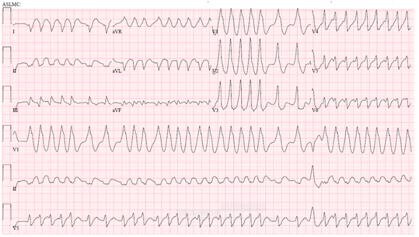
A 41 year old male with a rapid heart rate

 How would you treat this?

a. If the patient was haemodynamically stable?
b. If he was haemodynamically unstable?


From the American College of Cardiology Archives.

Monday, 4 January 2021

S-Gene Target Failure is Proxy for VOC 202012/01 COVID-19 strain

 The new COVID-19 variant, designated "Variant of Concern" or VOC 202012/01 by Public Health England, now accounts for the majority of transmission in the UK. The strain is characterised by the fact that the PCR used in most places in the UK- TaqPath by Thermo Fisher, has a dropout where the S gene should be. This is called S-Gene Target Failure, or simply SGTF.

The new variant has 23 mutations- 13 non-synonymous mutations, 4 deletions and  6 synonymous mutations- compared with the wild type variant, and one extra stop codon. Most of these mutations are in the gene for Spike protein, as shown below from the relevant PHE document.. One of these mutations, present at the N-terminal domain of the S-protein, is a deletion of 6 nucleotides- from 21765-21770, resulting in the loss of two amino acids- Histidine and Valine at position 69-70 of the Spike protein. It is the latter deletion that causes the characteristic SGTF on the TaqPath PCR. 

(Synonymous mutations lead to base pair substitutions which leave the amino acid unchanged, while non-synonymous mutations alter the coded amino acid)

TaqPath uses a 3-target PCR- it amplifies the S-gene, the N-gene (called N) and and Open Reading Frame called Orf1ab. COVID-19 has a further Orf called Orf8, which is not a target for TaqPath and therefore not amplified. (Open Reading Frames are DNA sequences without stop codons. When they are long, they are likely to code for protein, rather than being non-coding sequences).

For the VOC, the PCR cannot read the S-gene sequence due to deletion at 69-70, and so the S-gene "drops out", resulting in S-gene target failure. Only the other 2 targets, i.e. N and Orf1ab are amplified. 

Fully 99.7% of variants sequenced in the UK with SGTF belong to the new VOC strain. SGTF is thus used as a reliable proxy for the new strain.

However, as referred to elsewhere in this blog, the increased transmissibility of the new strain- between 40%-70% more than the wild type strain- is owed to another mutation in the Spike protein, namely N501Y, which affects the RBD, and is thought to increase the strength with which the S-protein binds to its cognate receptor on human cells- ACE2. 

Incidentally, the 69-70 deletion was seen in the Danish Cluster 5 variant, the one that led to a cull of millions of minks. Its clinical significance, unlike that of N501Y, is unknown.


Tuesday, 22 December 2020

Myxovirus Resistance Protein A (MxA) & Antibodies to Nuclear Matrix Protein-2 (NXP-2) in Dermatomyositis Sine Dermatitis

 There has long been debate on what is the most sensitive and specific marker that distinguishes Dermatomyositis (DM) from Polymyositis (PM) or Inclusion Body Myositis (IBM), and further whether anti-synthestase syndromes (ASS) should be included under DM. 

Where subjects present with a skin rash and muscle weakness with one of 5 DM specific autoantibodies, i.e. Mi-2, NXP-2, MDA-5, TIF-1gamma or SAE, the diagnosis of DM is straightforward. Given the difficulty in obtaining muscle biopsies within the NHS, particularly in DGHs, most Rheumatologists would settle for a diagnosis of DM in this scenario, particularly if the EMG too is characteristic of a myopathy.

But what if DM presents without a skin rash, as it can in approximately 8% of subjects. The distinction between DM one one hand and PM, IBM and ASS on the other is more than academic, firstly because of a higher association of cancer with the former, and secondly because of increasing reports of efficacy of JAK inhibitors for DM but not the other conditions.

In this situation, two tests can be useful. The first requires a muscle biopsy, and the second is included in the DM specific autoantibody panel.

First, the staining of the sarcoplasm of muscle for Myxovirus Resistance Protein A (MxA) is highly sensitive (76%) and 100% specific for DM. This is a Type I interferon induced protein and therefore an interferon signature. It is not seen in PM, IBM or ASS. Unlike the latter three, DM is an interferonopathy (which is why it responds to JAK inhibitors).

While other interferon signatures such as RIG-1 and ISG-15 are also quite specific for DM, as indeed are muscle biopsy findings of perifascicular atrophy (PFA) and deposition of membrane attack complex on capillaries, none of these are as sensitive as MxA. (While ASS also displays PFA on biopsy, the characteristic necrotic and regenerating fibres in perifascicular fibres sets it apart from the mainly degenerative fibres seen in DM).

The other useful marker of Dermatomyositis sine (without) dermatitis is the muscle specific antibody NXP-2. This is seen in fully 86% of subjects who have DM without skin involvement at presentation, but only in 28% of subjects who have DM with rash. Therefore, a subject presenting with myositis, but no rash and a positive NXP-2 should be treated as DM rather than PM. In a minority of such subjects, a typical rash may appear after many months or even years.

DM sine dermatitis should not be confused with amyopathic DM, which is characterised by rash and lung involvement without muscle involvement. Most such subjects are positive for MDA-5 and have a severe lung phenotype.

References

1. https://jamanetwork.com/journals/jamaneurology/fullarticle/2764337

2. https://pubmed.ncbi.nlm.nih.gov/30267437/

Sunday, 20 December 2020

The New VUI-202012/01 COVID-19 Variant Found in the United Kingdom

 The UK has just tightened its COVID tiers based on a fast spreading variant of the virus picked up by the COVID-19 Genomic Consortium. So far 1108 cases with this variant has been described. Apparently it is 70% more infectious than the prevalent D614G strain. Several European countries have imposed a summary bans on flights originating in the UK as a result.

It is fair to say that as a RNA virus, COVID-19 mutates continuously. A WHO analysis found that the rate of mutations for COVID-19 is 1.12 x 10^-3 mutations per site year, which is quite similar to the range of 0.80 x !0^-3 to 2.38 x 10^-3 per site year for the original SARS virus in 2003.

To put this into some context, the mutation rate in human beings is 1.2 x 10^-8 per generation. This translates to around 72 new mutations in a newborn. Mutagenesis is therefore an inevitable vicissitude of the genetic code.

You may be aware that the current circulating clade- D614G- originated in China in January 2020 and replaced the existing clade within 3 months. It is likely that VUI-202012/01 will become the dominant strain if allowed to spread.

Concern about emergence of new strains is reflected in the Danish government's recent decision to cull 17 million minks because they harboured a variant of the virus that was apparently not well neutralised by existing human antibodies.

The new variant VUI-202012/01 has 17 new mutations- the most important of which is N501Y involving the spike protein (which means that tyrosine has replaced asparagine in the 501st amino acid of the spike protein).

Looking at the DNA code, asparagine has 2 codons- AAC and AAT. Tyrosine also has two- TAC and TAT. This new strain is therefore due to A to T transversion at the first position of the putative codon. 

When a purine is replaced by a purine or pyrimidine by a pyrimidine, it is called a transition. Crossovers between purine and pyrimidine is called transversion. In general mutations caused by transition outnumber those due to transversions manyfold. The commonest mutations are due to C to T transition as cytosine bases are prone to be methylated at the 5'position and thence are spontaneously deaminated to thymine. Thus, the WHO database for COVID 19 in Feb 2020 showed 1670 C to T transitions compared with only 128 A to T transversions.

Thus, it is fair to say that the N501Y mutation in the VUI-202012/01 has persisted because it offers a survival advantage- i.e. infectivity, much as the G614 variant was more infective than the D614 variant. However, it must be said that there is no evidence that it is more dangerous. If anything, the D614G carried a lower mortality than the original strain, although this may have been due to better established treatment protocols.

Nor is there any evidence to suggest that the current vaccines will be less effective against the new strain. The current vaccines were in fact formulated against the original COVID strain and are just as effective against the D614G strain. This is due to the fact that protective antibodies target several epitopes and unless there is a significant conformational change in the shape of a protein, the vaccine generated antibodies will still neutralise.

I do think that this particular variant will take over despite the restrictions- just as D614G did. I also think there need be no undue cause for concern. With rapidly executed vaccination programmes, it should be possible to control the virus by spring.


Thursday, 17 December 2020

Getting The Most Out of Azathioprine

 Azathioprine is widely used in Rheumatology for conditions such as vasculitis and Lupus, and by some in Rheumatoid. It certainly has a place in gastroenterology for the management of ulcerative colitis and Crohn's disease, where it is more effective for maintenance of remission than 5-ASA.

However, the use of Azathioprine is beset with problems. Around 10% of subjects have hypersensitivity reactions to this drug, comprising fever, nausea and diarrhoea, fatigue, malaise and myalgia, mandating rapid discontinuation. A further 25-30% have side effects such as hepatotoxicity in the form of transaminitis or myelotoxicity manifesting as neutropenia. As a result, fully 40% of subjects that start azathioprine do not continue with the drug.

Observance of some simple principles can obviate these difficulties. The first of these is widely practiced- measuring TPMT levels before starting azathioprine. In the general population, 89% of subjects are homozygous for high metabolism of azathioprine (high TPMT) , 11% are heterozygous and 0.3% have low levels of TPMT. 

It is common practice not to use azathioprine in subjects with lowish TPMT- these are the heterozygotes. This is a missed opportunity as these are the very subjects who are likely to respond the best to the drug. Thus, people with TPMT levels >25 U/ml need no dose adjustment. (Below 25 U/ml, halve the dose, and monitor more frequently). Conversely, those with levels above 65 U/ml, although reassuring on the face of it, are likely to have treatment failure.

Azathioprine is a prodrug of 6-MP. Most of the ingested azathioprine is non-enzymatically cleaved to 6-MP in the liver. Yet, there is a marked non-familiarity with 6-MP amongst Rheumatologists and perhaps to a lesser extent among Gastroenterologists. Several studies show that in subjects with hypersensitivity to azathioprine, nearly 70% tolerate a switch to 6-MP. The dose for 6-MP is half that of azathioprine (1-1.5 mg/kg body weight, rather than 2-2.5 mg/kg). Subjects with flu like reactions, nausea, emesis, myalgias and arthralgias on Azathioprine are likely to be able to switch successfully to 6-MP. OTOH, those with hepatotoxicity and pancreatitis are likely to have the same problems with 6-MP.

The converse does not apply. Thus, subjects who are intolerant to 6-MP should not be switched to azathioprine.

While most practitioners are aware of the importance of measuring TPMT before commencing thiopurines (azathioprine or 6-MP) , there is less awareness of the rather high usefulness of measuring thiopurine metabolites. Two metabolites are measured by most labs- 6-Thioguanine nucleotide (6-TGN) and 6-Methyl mercaptopurine (6-MMPN). The metabolite 6-MMPN is produced en-route to 6-TGN (please see diagram). It is the blood level of 6-TGN that indicates efficacy of azathioprine. Within a blood 6-TGN range of 235-450 pmol/8 x 10^8 RBC (send whole blood in an EDTA tube, just like TPMT), azathioprine is likely to be effective. Below 235, efficacy is likely to be low. This latter could be due to 2 reasons- non compliance, or the fact that not enough azathioprine is being metabolised to the active metabolite 6-TGN. In the latter case , blood 6-MMPN levels will be high (range 0-5700). 

The combination of low 6-TGN and high 6-MMPN levels presages treatment failure and hepatotoxicity, and is described as azathioprine resistance. Unlike with non-compliance (where 6-TGN is low and 6-MMPN is normal), increasing the dose of azathioprine where resistance exists is only likely to lead to hepatotoxicity without increasing efficacy.

Keep in mind though that unlike hepatotoxicity, isolated neutropenia is a surrogate marker for the effectiveness of azathioprine. Here, the 6-TGN levels are likely to be high, and may require a reduction in dosage, rather than discontinuation, as with transaminitis. (If hepatotoxicity and neutropenia occur together, discontinue the drug).

There are anecdotal reports that in subjects with high 6-MMPN levels, splitting the dose of azathioprine is likely to improve efficacy and reduce toxicity, while lowering the level of 6-MMPN and maintaining that of 6-TGN. However, this is based on observations from a single study.

It is fair to say that we could be using azathioprine a lot more effectively than we currently do.


 Figure. Thiopurine metabolic pathway. Metabolic pathway for AZA and 6MP is shown in the diagram. AZA: Azathioprine; 6-MP: 6-mercaptopurine; 6-TU: Thiouric acid; 6-MMP: 6-methylmercaptopurine; TIMT: Thiopurine methyl-transferase; 6-MMPR: Methyl-mercaptopurine ribonucleotide; TXMP: 6-thioxanthosine monophosphate; 6-TGN: Thioguanine nucleotide; 6-TG: Thioguanine; 6-TGDP: 6-thioguanine diphosphate; 6-TGTP: 6-thioguanine triphosphate; XO: Xanthine oxidase; TPMT: Thiopurine methyltransferase; HPRT: Hypoxanthine phosphoribosyl transferase.


Reference:

https://www.ncbi.nlm.nih.gov/pmc/articles/PMC3208360/


Sunday, 13 December 2020

The Purinergic Pathway in Inflammation, Heart Disease & Cancer

 So you drink a cup of coffee because you are tired. It relieves your fatigue and headache? How does it do it? 

Or for that matter, how do methotexate and sulfasalazine relieve inflammation in rheumatoid arthritis or inflammatory bowel disease? How do clopidogrel and dipyridamole work? And what are the most important cellular markers of effective adoptive cell transfer therapy?

The answer to all these questions lies in the all-important purinergic system. This is the dance between the nucleotides ATP and ADP on one hand and adenosine on the other. They often have diametrically opposite effects in health and disease.

ATP is of course the currency of energy in eukaryotic cells. However, here we are referring to extracellular ATP, acting in a paracrine fashion on specific receptors. Such extracellular ATP may be released from necrotic cells, or leave apoptotic cells through pannexin channels or exit inflammatory cells such as neutrophils through connexin channels- connexin 37 & 44, specifically. They can also be part of vesicles, released from cells through exocytosis.

Whatever the origin of ATP, or the closely related dinucleotide ADP, formed from ATP, they act through two groups of cell surface receptors- the first, called P2Y receptors, are metabotropic receptors, i.e. they directly respond to the nucleotides- these are G-protein coupled receptors, and the second, called P2X receptors, are ionotropic receptors, i.e. ligand gated ion channels that open in response to inward flux of calcium (mostly) or sodium, or outward flux of potassium.

On the other side of the spectrum, sit P1 receptors, which ligate extracellular adenosine. These are also GPCRs. There are 4 types, adenosine receptor A1 (also called ADORA1), A2A(ADORA2A), A2B(ADORA2B) and A3(ADORA3). Of these, A2A and A2B are coupled to Gs and lead to increased levels of cellular cAMP, resulting in profound immunosuppression. A1 & A3 inhibit the formation of cAMP through Gi/o and are therefore generally immune promoting. While A1, A2A and A3 are high affinity adenosine receptors, A2B has low affinity, and is only stimulated under pathologic conditions such as high prevailing levels of adenosine in a hypoxic tumour microenvironment.

How is adenosine formed extracellularly? It is principally formed by the action of 2 sequential cell surface enzymes called CD39 and CD73. CD39 is an ectonucleoside triphosphate diphosphohydrolase, which converts ATP and ADP to AMP. CD73 is an ecto-5'-nuleotidase, that converts AMP to adenosine.

Adenosine can sometimes be generated by other enzymes from ATP & AMP, namely alkaline phosphatase, which has been described as a "promiscuous" enzyme.

Extracellular adenosine is short lived and pushed inside the cell through a couple of channels called Equilibrative Nucleoside Transporters 1&2 , also called ENT1 & ENT2.

Extracellular ATP is pro-inflammatory. It activates the NLRPR3 inflammasome in neutrophils and monocytes. The resulting Caspase1 cleaves pro-IL1 and pro-IL18 into their active forms. 

Adenosine, on the other hand is anti-inflammatory. It reduces inflammation through the A2A receptors present on neutrophils and lymphocytes, by increasing levels of cAMP. Remember, A2A is a high affinity receptor for adenosine.

Methotrexate and sulfasalazine owe their anti-inflammatory effect at least partly due to the fact that they stimulate CD73, which increases the formation of extracellular adenosine from AMP.

What of the A1, A2B and A3 receptors for adenosine? These have all been exploited pharmacologically. The heart blocking effect of adenosine in terminating SVT is exerted through the A1 receptor, while its vasodilating effect in cardiac stress testing is due to its action on A2B receptor on vascular endothelial cells. Stimulation of A3 receptors in non-pigmented cells in the anterior chamber of the eye leads to increased production of aqueous humour, and can be useful for treating sicca symptoms.

Dipyridamole inhibits the ENT1 & 2 channels, thus leading to increased levels of  extracellular adenosine. Hence its use in pharmacological cardiac stress testing.

As adenosine is formed from ATP and ADP, their levels vary inversely with each other. Thus, in inflammatory bowel disease, tissue hypoxia leads to increased production of HIF, which, acting as a transcription factor, stabilises the promoters for CD73 and A2B. A similar transcription factor called Sp1 binds to and stabilises the promoter for CD39. The net result is an increase in extracellular adenosine and a reduction in ATP. This leads to reduction in inflammation, both due to a fall in extracellular ATP levels and stimulation of A2A receptors on neutrophils and lymphocytes by adenosine. The latter also stimulates A2B receptors and maintains epithelial integrity, presumably by promoting healing through augmented blood flow.

The purinergic system, in particular ADP, plays an important role in the function of platelets. Thus, ADP stimulates P2Y1 receptors on platelets and through the G-protein Gq, activates phospholipase C. The downstream effect of this is change in the shape of platelets through the actin cytoskeleton. Similarly ADP activates the P2Y12 receptor, which, through the intermediation of the Gi G-protein switches off adenylyl cyclase, decreases cAMP and activates the GPIIa/IIIb receptor, thus facilitating the binding of platelets to fibrinogen, resulting in platelet aggregation.

Clopidogrel is a P2Y12 inhibitor. It is a prodrug and needs to be activated in the liver. This particular stage can be affected in certain mutations and thus reduce the efficacy of clopidogrel in affected subjects.

Stimulation of A2A and A2B receptors leads to platelet inhibition, explaining the efficacy of dipyridamole in prevention of ischaemic stroke.

While extracellular adenosine is regarded as a safety signal in ischaemia and reperfusion, where it reduces inflammation and tissue damage, it can have the opposite effect in cancer. In general, while its immunosuppressive effect on T-lymphocytes reduces autoimmunity, it can be a hindrance in fighting cancerous cells. In a recent paper in Nature, the investigators found that the subset of cancer sufferers who had the highest benefit from adoptive T cell therapy had a higher proportion of CD8+CD39-CD69- T-cells in the infusate. It is possible that increased expression of CD39 on T-cells leads to production of extracellular adenosine, immunosuppression and T-cell exhaustion, although this is yet to be confirmed. In general, adenosine is found in higher quantities in hypoxic tumour microenvironments, although this may be consequence rather than cause of tumour survival.

Finally, to the salutary effects of that cup of coffee. It is thought that caffeine reduces fatigue by inhibiting cerebral adenosine A2A receptors.

Wednesday, 25 November 2020

The Science Behind DNA Analysis of Archaeological Human Remains

 DNA, even when well preserved, will not last for more than a few hundred thousand years. Therefore the description of DNA sequences in million year old dinosaur remains is due to contamination from modern ambient DNA, which is widespread in labs and elsewhere. The PCR will amplify any DNA, modern or prehistoric.

There are however certain clues to the age of DNA remains. The first is fragmentation. Ancient DNA fragments are between 50 and 500 bases long due to degradation. In fact most are no longer than 100 nucleotides.

Second, and most reliable is the observation that at both ends of DNA (5' and 3'), Cytosine (C) is deaminated to Uracil (U). This is an effect that occurs after death due to bacterial cytosine deaminase. This latter enzyme is not present in mammals (not to be confused with AID or activation induced cytidine deaminase, which is present during development and plays a major role in the affinity maturation and somatic hypermutation of B cells, in effect giving B cells their antigen specificity).

Thus if an archaeological remains has a high proportion of uracil bases in DNA- remember, this base is only present in RNA, it indicates that the specimen is thousands of years old. Archaeologists use an enzyme called uracil-DNA- glycosylase (UDG) to break the bond between deoxyribose sugar and uracil and remove the uracil bases. 

Not doing so leads to a curious phenomenon. The PCR used to amplify the DNA remains reads the uracil as thymine (T), since thymine is the native DNA base, not uracil. Hence, on the supplementary (anti-sense) strand, it puts an adenine in place of the guanine (which paired with the original cytosine). The uracil on the sense strand is now replaced with thymine to pair with adenine. The original C-G pairing has therefore now been replaced by an A-T pair. Thus there is miscoding caused by the PCR process manifesting as "transition " of C to T and G to A. (In contrast, when a purine is replaced by pyrimidine base, it is described as "transversion"). 

There can be blocking lesions caused by the formation of non-physiological chemical bonds between say DNA & protein, which can make it difficult for PCR to read through the ancient DNA. This is difficult to overcome.

Archaeologists have databases comprising thousands of single nucleotide polymorphisms (SNPs) mapped from certain lineages such as Armenian farmers or Iranian invaders which they then compare with the putative SNPs from the sequenced DNA sample to see if there is a match.

Here, mitochondrial DNA, which is of course maternal derived (the sperm does not contain mitochondria) is more useful than nuclear DNA in lineage tracing. Although mitochondrial DNA shares the same predisposition for degradation with time elapsed since death as nuclear DNA, there are far more copies of a given DNA sequence in mitochondria than in chromosomes. Thus, while the nucleus will only have two copies of a given allele (on the 2 chromosomes), each mitochondrion has 10-100 copies- a phenomenon known as heteroplasmy, which explains why mitochondrial mutations cause such variable phenotypes unlike mutations that occur in chromosomes. Mitochondrial DNA therefore is favoured by archaeologists for lineage tracing.

Are there any other ways to determine where a long dead person originated, apart from DNA sequencing? As it happens, there is.

The isotopes of Strontium which are deposited in bone and teeth (dentine and enamel) during development are a reflection of the area where the person grew up in. There are 4 isotopes of Strontium- Sr84, Sr86, Sr87 and Sr88. Sr 87 is not a natural isotope. It is formed by beta decay from Rubidium 87. The rocks and water and therefore, by extension, the remains of a person or animal growing up in a certain area will always display a specific Sr87:Sr88 ratio that is native to that area. When that person migrates to a different geographical area, his teeth, which are often well preserved thousands of years after death, will still carry the signature Sr87:Sr88 ratio of the area where he or she grew up in , as the signature is established in the developing teeth or bones, and does not change after death. 

It is thus possible to say that the woman's remains found in a Harappan valley, in fact grew up in Iran.


Saturday, 14 November 2020

Paradoxical Effect of Caesin Kinase Inhibition in Deletion 5q Myelodysplastic Syndrome

 Caesin Kinase (CK1) is common to the Wnt-beta catenin pathway and the Hedgehog pathway. Along with other kinases, it acts as an inhibitor in both pathways to reduce the transcription of genes in response to wnt and hedgehog proteins respectively. 

Myelodysplastic syndrome (MDS) due to deletion of 5q (5q del) has an unique phenotype which can be explained by the differential action of CK1 on the wnt-beta catenin pathway. 

Subjects with 5q del have macrocytic anaemia, mild thrombocytosis with dysplastic, hypolobated megakaryocytes. MDS in 5q del is uniquely responsive to immunomodulators such as lenalidomide, although the anaemia does relapse after 2-3 years due to new mutations in other genes such as RUNX1.

Deletion of 5q leads to haploinsufficiency of the CK1A1 gene located on 5q.32. Lenalidomide inhibits the remaining CK1A1 allele and reverses the phenotype of MDS. This is admittedly non-intuitive, as there seems to be an abrogation of the dose-response effect here. If haploinsufficiency leads to a phenotype, how can suppressing the remaining normal allele reverse that phenotype (rather than worsen it)?

This apparent paradox is explained by the effect of CK1 on the wnt-beta catenin pathway. Haploinsufficiency of CK1 removes some of the inhibition exercised on the wnt-beta catenin pathway. The latter leads to enhanced survival and proliferation of the neoplastic clone, causing MDS.

However, genetic knockdown in mice or pharmacological inhibition of the remaining CK1A1 allele by lenalidomide in 5q del MDS sufferers leads to complete disinhibition of the wnt-beta catenin pathway. While this initially stimulates the haemopoetic progenitors of the neoplastic clone, continued stimulation soon leads to stem cell exhaustion and death of the progenitor cells now bereft of any CK1 activity.


Wednesday, 11 November 2020

Reversal of The Earth's Magnetic Axis

 The magnetic axis of celestial objects flips periodically, including a complete reversal (by 180 degrees). For example, the sun's magnetic axis reverses direction every 11 years. However, the Earth's geomagnetic axis flips much less often. The last time it reversed completely was 780,000 years ago, a phenomenon called the Matuyama-Bunhes reversal after the two scientists who described it. Since then, the Earth's magnetic field has tried to flip on 10 occasions, but on each occasion it has reverted back to its current axis.

It's fair to say therefore that such reversals happen very infrequently for the Earth. When it does happen though, it affects the polarity of magnetic material in lava flows, sea beds etc which can be detected in rocks and fossils. 

The Curie temperature is one above which magnetic substances (which includes all minerals containing iron, nickel and cobalt) lose their magnetic properties. This varies between 580 and 680 degrees Celsius for the oxides of iron -Fe2O3 and Fe3O4. Conversely, when cooled below this temperature, such objects regain their magnetism. Thus, igneous rocks have inherent magnetism dating back to the time when they were formed from cooling lava flows millions of years ago.

The reversal of Earth's magnetic axis is not instantaneous- it occurs slowly- over thousands of years. During this period, magnetic material in cooling magma will take up the polarity of the reversed magnetic polarity of Earth, and the rocks formed therefrom would reflect this reversed polarity for ever. Thus, geologists can analyse such rocks, or fossils which have enclosed magnetic material in sea beds and make a fair guess as to their age. This is one way of fossil or rock dating.